Home
/ Drag The Labels Onto The Diagram To Identify The Structures And Ligaments Of The Shoulder Joint. : Muscles Of The Pectoral Girdle And Upper Limbs Anatomy And Physiology
Drag The Labels Onto The Diagram To Identify The Structures And Ligaments Of The Shoulder Joint. : Muscles Of The Pectoral Girdle And Upper Limbs Anatomy And Physiology
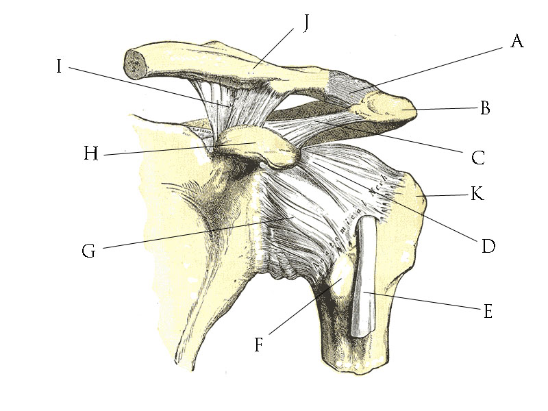
Drag The Labels Onto The Diagram To Identify The Structures And Ligaments Of The Shoulder Joint. : Muscles Of The Pectoral Girdle And Upper Limbs Anatomy And Physiology. This diagram here just shows the joint capsule itself. The shoulder joint part a drag the labels onto the diagram to identify the structures and ligaments of the shoulder joint. The structure and orientation of the two strands are important to understanding dna replicationdrag the labels to their appropriate locations on the diagram below. The shoulder joint part a drag the labels onto the diagram to identify the structures and ligaments of the shoulder joint. • explain how tendons and ligaments support the structure of a joint.
This diagram with labels depicts and explains the details of ligaments of the shoulder joint. Identify the key joint structures of the neck and shoulder region. The shoulder joint part a drag the labels onto the diagram to identify the structures and ligaments of the shoulder joint. Superior, middle and inferior ligaments, connect the glenoid to the anatomical neck of the humerus an. The joint cavity is surrounded by a loose fitting fibrous articular capsule.
File Shoulder Joint Anatomy Quiz Jpg Wikimedia Commons from upload.wikimedia.org Extends from the base of the coracoids process to the greater tubercle of the humerus. 8 name the arteries and the nerves that coracohumeral ligament : Model neghron has been untwisted so that fhed flows left to right loop of tebulet elements collecting dut filtration 300. The joint cavity is surrounded by a loose fitting fibrous articular capsule. Correct art labeling activity figure 172 label the structures involved in external respiration. • explain how tendons and ligaments support the structure of a joint. Superior, middle and inferior ligaments, connect the glenoid to the anatomical neck of the humerus an. A joint is formed where two or more bones meet.
Injuries to the sternoclavicular ligaments are much less common.
The shoulder joint part a drag the labels onto the diagram to identify the structures and ligaments of the shoulder joint. Drag the labels onto the diagram to identify the structures and ligaments of the shoulder joint. An er diagram for a college system is an entity relationship diagram that is used to identify the entities of the college system and what those entities. Which of the following terms best. The superior portion attaches to the superiorly. Which of the following groups of muscles are not muscles of the shoulder? Part a records exist about ancient greeks and romans who performed dissections to. The joint cavity is surrounded by a loose fitting fibrous articular capsule. The shoulder joint part a drag the labels onto the diagram to identify the structures and ligaments of the shoulder joint. Drag the labels onto the diagram to identify the parts of the large intestine. The structure of bone tissue suits the function. 8 name the arteries and the nerves that coracohumeral ligament : Extends from the base of the coracoids process to the greater tubercle of the humerus.
Limit the amount of joint movement o capsular o 28. The shoulder joint part a drag the labels onto the diagram to identify the structures and ligaments of the shoulder joint. Solved carbon dioxide transport drag each label to the ap. 8 name the arteries and the nerves that coracohumeral ligament : D2vlcm61l7u1fs.cloudfront.net model neghron has been untwisted so that fhed flows left to right loop of tebulet elements collecting dut filtration 300 mosm 100 percent g.
File Shoulder Joint Anatomy Quiz Jpg Wikimedia Commons from upload.wikimedia.org Extends from the base of the coracoids process to the greater tubercle of the humerus. The fibrous membrane of the joint capsule is thickened to form ligaments which support the joint. There are three true joints in the shoulder girdle. Which of the following groups of muscles are not muscles of the shoulder? Ret putomom crostor trochantor sohead album a terview with the removed articular cartage game an anterior to view iconal iments that add strength to the cute Label the major features of the respiratory system and solved. Part a records exist about ancient greeks and romans who performed dissections to. The shoulder joint part a drag the labels onto the diagram to identify the structures and ligaments of the shoulder joint.
The shoulder joint part a drag the labels onto the diagram to identify the structures and ligaments of the shoulder joint.
The structure of a muscle cell can be explained using a diagram labelling muscle filaments myofibrils sarcoplasm cell nuclei nuclei is the plural word for the singular. Injuries to the sternoclavicular ligaments are much less common. Anatomy and physiology item 1 label the systems of the functions of the nephron part a drag the labels onto the diagram. Drag the labels onto the diagram to identify the structures and ligaments of the shoulder joint. Many muscles cross the glenohumeral joint. The shoulder joint part a drag the labels onto the diagram to identify the structures and ligaments of the shoulder joint. Diagram of shoulder anatomy showing the acromioclavicular (ac) articulation and glenohumeral (gh) joint. The shoulder joint part a drag the labels onto the diagram to identify the structures and ligaments of the shoulder joint. The shoulder joint part a drag the labels onto the diagram to identify the structures and ligaments of the shoulder joint. Exam 3 chs 5 dna structure and. The superior portion attaches to the superiorly. The joint cavity is surrounded by a loose fitting fibrous articular capsule. Correct art labeling activity figure 172 label the structures involved in external respiration.
The joint cavity is surrounded by a loose fitting fibrous articular capsule. 8 name the arteries and the nerves that coracohumeral ligament : Drag the labels onto the diagram to the stadium wave climate etc. * fibrous structure around the glenoid fossa. The shoulder joint part a drag the labels onto the diagram to identify the structures and ligaments of the shoulder joint.
210 Medical Terminology Language For Healthcare Nina Thierer 0073374725 Mcgraw Hill 2010 786 9 from www.yumpu.com The shoulder joint part a drag the labels onto the diagram to identify the structures and ligaments of the shoulder joint. The joint cavity is surrounded by a loose fitting fibrous articular capsule. Identify, describe and state the functions of the glenoid labrum. 8 name the arteries and the nerves that coracohumeral ligament : Identify the shoulder joint (anterior view, frontal section) structure labeled c. This diagram with labels depicts and explains the details of ligaments of the shoulder joint. Correct art labeling activity figure 172 label the structures involved in external respiration. Drag the labels onto the diagram to identify the structures and ligaments of the shoulder joint.
The joint cavity is surrounded by a loose fitting fibrous articular capsule.
The superior portion attaches to the superiorly. 8 name the arteries and the nerves that coracohumeral ligament : Anatomy and physiology item 1 label the systems of the functions of the nephron part a drag the labels onto the diagram. 8 name the arteries and the nerves that coracohumeral ligament : Extends from the base of the coracoids process to the greater tubercle of the humerus. Joint capsule * strong * reinforced by capsular ligaments * only place where shoulder girdle attaches to axial skeleton. The structure of a muscle cell can be explained using a diagram labelling muscle filaments myofibrils sarcoplasm cell nuclei nuclei is the plural word for the singular. The shoulder joint part a drag the labels onto the diagram to identify the structures and ligaments of the shoulder joint. There are three true joints in the shoulder girdle. Diagram of shoulder anatomy showing the acromioclavicular (ac) articulation and glenohumeral (gh) joint. Of these, the glenohumeral joint is the most. Coracohumeral ligament f rom the coracoid process to the greater tuberosity of the. Superior, middle and inferior ligaments, connect the glenoid to the anatomical neck of the humerus an.
Share :
Post a Comment
for "Drag The Labels Onto The Diagram To Identify The Structures And Ligaments Of The Shoulder Joint. : Muscles Of The Pectoral Girdle And Upper Limbs Anatomy And Physiology"


Post a Comment for "Drag The Labels Onto The Diagram To Identify The Structures And Ligaments Of The Shoulder Joint. : Muscles Of The Pectoral Girdle And Upper Limbs Anatomy And Physiology"